|
 |
 | We have recently changed our website. Looking for the previous site? |
Abstracts of Articles in 2017
To request any journal reprints, please contact us.- Gaussian Accelerated Molecular Dynamics in NAMD
- NMR Structure-Based Optimization of Staphylococcus aureus Sortase A Pyridazinone Inhibitors
- Spectroscopic and Computational Investigations of Ligand Binding to IspH: Discovery of Non-Diphosphate Inhibitor
- Activation Mechanisms of the Sphingosine-1-Phosphate Receptor
- Effect of Donor Atom Identity on Metal Binding Pharmacophore Coordination
- CRISPR-Cas9 conformational activation as elucidated from enhanced molecular simulations
- Gaussian Accelerated Molecular Dynamics: Theory, Implementation and Applications
- ‘MARTINIZING’ the Variational Implicit Solvent Model (VISM): Solvation Free Energy for Coarse-Grained Proteins
- Protospacer Adjacent Motif-Induced Allostery Activates CRISPR-Cas9
- Manipulating Protein–Protein Interactions in Nonribosomal Peptide Synthetase Type II Peptidyl Carrier Proteins
Gaussian Accelerated Molecular Dynamics in NAMDJ. Chem. Theory Comput., 13 (1), pp9-19 (2017)
Gaussian accelerated molecular dynamics (GaMD) is a recently developed enhanced sampling technique that provides efficient free energy calculations of biomolecules. Like the previous accelerated molecular dynamics (aMD), GaMD allows for "unconstrained" enhanced sampling without the need to set predefined collective variables and so is useful for studying complex biomolecular conformational changes such as protein folding and ligand binding. Furthermore, because the boost potential is constructed using a harmonic function that follows Gaussian distribution in GaMD, cumulant expansion to the second order can be applied to recover the original free energy profiles of proteins and other large biomolecules, which solves a long-standing energetic reweighting problem of the previous aMD method. Taken together, GaMD offers major advantages for both unconstrained enhanced sampling and free energy calculations of large biomolecules. Here, we have implemented GaMD in the NAMD package on top of the existing aMD feature and validated it on three model systems: alanine dipeptide, the chignolin fast-folding protein, and the M muscarinic G protein-coupled receptor (GPCR). For alanine dipeptide, while conventional molecular dynamics (cMD) simulations performed for 30 ns are poorly converged, GaMD simulations of the same length yield free energy profiles that agree quantitatively with those of 1000 ns cMD simulation. Further GaMD simulations have captured folding of the chignolin and binding of the acetylcholine (ACh) endogenous agonist to the M muscarinic receptor. The reweighted free energy profiles are used to characterize the protein folding and ligand binding pathways quantitatively. GaMD implemented in the scalable NAMD is widely applicable to enhanced sampling and free energy calculations of large biomolecules.
Gaussian accelerated molecular dynamics (GaMD) is a recently developed enhanced sampling technique that provides efficient free energy calculations of biomolecules. Like the previous accelerated molecular dynamics (aMD), GaMD allows for "unconstrained" enhanced sampling without the need to set predefined collective variables and so is useful for studying complex biomolecular conformational changes such as protein folding and ligand binding. Furthermore, because the boost potential is constructed using a harmonic function that follows Gaussian distribution in GaMD, cumulant expansion to the second order can be applied to recover the original free energy profiles of proteins and other large biomolecules, which solves a long-standing energetic reweighting problem of the previous aMD method. Taken together, GaMD offers major advantages for both unconstrained enhanced sampling and free energy calculations of large biomolecules. Here, we have implemented GaMD in the NAMD package on top of the existing aMD feature and validated it on three model systems: alanine dipeptide, the chignolin fast-folding protein, and the M
NMR Structure-Based Optimization of Staphylococcus aureus Sortase A Pyridazinone InhibitorsChem. Biol. Drug Design 90, 327–344 (2017)
Staphylococcus aureus is a leading cause of hospital-acquired infections in the USA and is a major health concern as methicillin-resistant S. aureus and other antibiotic-resistant strains are common. Compounds that inhibit the S. aureus sortase (SrtA) cysteine transpeptidase may function as potent anti-infective agents as this enzyme attaches virulence factors to the bacterial cell wall. While a variety of SrtA inhibitors have been discovered, the vast majority of these small molecules have not been optimized using structure-based approaches. Here we have used NMR spectroscopy to determine the molecular basis through which pyridazinone-based small molecules inhibit SrtA. These inhibitors covalently modify the active cysteine thiol and partially mimic the natural substrate of SrtA by inducing the closure of an active site loop. Computational and synthetic chemistry methods led to second-generation analogues that are ~70-fold more potent than the lead molecule. These optimized molecules exhibit broad-spectrum activity against other types of class A sortases, have reduced cytotoxicity, and impair SrtA-mediated protein display on S. aureus cell surface. Our work shows that pyridazinone analogues are attractive candidates for further development into anti-infective agents, and highlights the utility of employing NMR spectroscopy and solubility-optimized small molecules in structure-based drug discovery.
Staphylococcus aureus is a leading cause of hospital-acquired infections in the USA and is a major health concern as methicillin-resistant S. aureus and other antibiotic-resistant strains are common. Compounds that inhibit the S. aureus sortase (SrtA) cysteine transpeptidase may function as potent anti-infective agents as this enzyme attaches virulence factors to the bacterial cell wall. While a variety of SrtA inhibitors have been discovered, the vast majority of these small molecules have not been optimized using structure-based approaches. Here we have used NMR spectroscopy to determine the molecular basis through which pyridazinone-based small molecules inhibit SrtA. These inhibitors covalently modify the active cysteine thiol and partially mimic the natural substrate of SrtA by inducing the closure of an active site loop. Computational and synthetic chemistry methods led to second-generation analogues that are ~70-fold more potent than the lead molecule. These optimized molecules exhibit broad-spectrum activity against other types of class A sortases, have reduced cytotoxicity, and impair SrtA-mediated protein display on S. aureus cell surface. Our work shows that pyridazinone analogues are attractive candidates for further development into anti-infective agents, and highlights the utility of employing NMR spectroscopy and solubility-optimized small molecules in structure-based drug discovery.
Spectroscopic and Computational Investigations of Ligand Binding to IspH: Discovery of Non-Diphosphate InhibitorChemBio Chem, Vol. 8 (10), pp 914-920 (2017)
Isoprenoid biosynthesis is an important area for anti-infective drug development and one target is IspH, (E)-1-hydroxy-2-methyl-but-2-enyl 4-diphosphate (HMBPP) reductase, which forms isopentenyl diphosphate and dimethylallyl diphosphate from HMBPP in a 2H+/2e- reduction. IspH contains a 4Fe-4S cluster and here, we first investigated how small molecules can bind to the cluster using HYSCORE and NRVS spectroscopies. The results of these as well as other structural and spectroscopic investigations led to the conclusion that in most cases, ligands bind to IspH 4Fe-4S clusters via h1 coordination, forming tetrahedral geometries at the unique 4th Fe, ligand side-chains preventing further ligand (e.g. H2O, O2) binding. Based on these ideas, we sought using in silico methods to find drug-like inhibitors that might occupy the HMBPP substrate binding pocket and bind to Fe, leading to the discovery of a barbituric acid analog having a Ki ~ 500 nM against Pseudomonas aeruginosa IspH.
Isoprenoid biosynthesis is an important area for anti-infective drug development and one target is IspH, (E)-1-hydroxy-2-methyl-but-2-enyl 4-diphosphate (HMBPP) reductase, which forms isopentenyl diphosphate and dimethylallyl diphosphate from HMBPP in a 2H+/2e- reduction. IspH contains a 4Fe-4S cluster and here, we first investigated how small molecules can bind to the cluster using HYSCORE and NRVS spectroscopies. The results of these as well as other structural and spectroscopic investigations led to the conclusion that in most cases, ligands bind to IspH 4Fe-4S clusters via h1 coordination, forming tetrahedral geometries at the unique 4th Fe, ligand side-chains preventing further ligand (e.g. H2O, O2) binding. Based on these ideas, we sought using in silico methods to find drug-like inhibitors that might occupy the HMBPP substrate binding pocket and bind to Fe, leading to the discovery of a barbituric acid analog having a Ki ~ 500 nM against Pseudomonas aeruginosa IspH.
Activation Mechanisms of the Sphingosine-1-Phosphate ReceptorProtein Science, Vol. 26, (6), pp. 1150-1160 (2017)
Activation of the first sphingosine-1-phosphate receptor (S1PR1) promotes permeability of the blood brain barrier, astrocyte and neuronal protection, and lymphocyte egress from secondary lymphoid tissues. Although an agonist often activates the S1PR1, the receptor exhibits high levels of basal activity. In this study, we performed long-timescale molecular dynamics and accelerated molecular dynamics (aMD) simulations to investigate activation mechanisms of the ligand-free (apo) S1PR1. In the aMD enhanced sampling simulations, we observed four independent events of activation, which is characterized by close interaction between Y3117.53 and Y2215.58 and increased distance between the intracellular ends of transmembrane (TM) helices 3 and 6. Although transmembrane helices TM3, TM6, TM5 and, TM7 are associated with GPCR activation, we discovered that their movements are not necessarily correlated during activation. Instead, TM5 showed a decreased correlation with each of these regions during activation. During activation of the apo receptor, Y2215.58 and Y3117.53 became more solvated, because a water channel formed in the intracellular pocket. Additionally, a lipid molecule repeatedly entered the receptor between the extracellular ends of TM1 and TM7, providing important insights into the pathway of ligand entry into the S1PR1. This article is protected by copyright. All rights reserved.
Activation of the first sphingosine-1-phosphate receptor (S1PR1) promotes permeability of the blood brain barrier, astrocyte and neuronal protection, and lymphocyte egress from secondary lymphoid tissues. Although an agonist often activates the S1PR1, the receptor exhibits high levels of basal activity. In this study, we performed long-timescale molecular dynamics and accelerated molecular dynamics (aMD) simulations to investigate activation mechanisms of the ligand-free (apo) S1PR1. In the aMD enhanced sampling simulations, we observed four independent events of activation, which is characterized by close interaction between Y3117.53 and Y2215.58 and increased distance between the intracellular ends of transmembrane (TM) helices 3 and 6. Although transmembrane helices TM3, TM6, TM5 and, TM7 are associated with GPCR activation, we discovered that their movements are not necessarily correlated during activation. Instead, TM5 showed a decreased correlation with each of these regions during activation. During activation of the apo receptor, Y2215.58 and Y3117.53 became more solvated, because a water channel formed in the intracellular pocket. Additionally, a lipid molecule repeatedly entered the receptor between the extracellular ends of TM1 and TM7, providing important insights into the pathway of ligand entry into the S1PR1. This article is protected by copyright. All rights reserved.
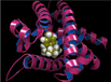
Effect of Donor Atom Identity on Metal Binding Pharmacophore CoordinationJ. Biol. Inorg. Chem. 22, 605–613 (2017)
The inhibition and binding of three metal-binding pharmacophores (MBPs), 2-hydroxycyclohepta-2,4,6-trien-1-one (tropolone), 2-mercaptopyridine-N-oxide (1,2-HOPTO), and 2-hydroxycyclohepta-2,4,6-triene-1-thione (thiotropolone) to human carbonic anhydrase II (hCAII) and a mutant protein hCAII L198G were investigated. These MBPs displayed bidentate coordination to the active site Zn(II) metal ion, but the MBPs respond to the mutation of L198G differently, as characterized by inhibition activity assays and X-ray crystallography. The L198G mutation increases the active site volume thereby decreasing the steric pressure exerted on MBPs upon binding, allowing changes in MBP coordination to be observed. When comparing the binding mode of tropolone to thiotropolone or 1,2-HOPTO (O,O versus O,S donor sets), structural modifications of the hCAII active site were shown to have a stronger effect on MBPs with an O,O versus O,S donor set. These findings were corroborated with density functional theory (DFT) calculations of model coordination complexes. These results suggest that the MBP binding geometry is a malleable interaction, particularly for certain ligands, and that the identity of the donor atoms influences the response of the ligand to changes in the protein active site environment. Understanding underlying interactions between a MBP and a metalloenzyme active site may aid in the design and development of potent metalloenzyme inhibitors.
The inhibition and binding of three metal-binding pharmacophores (MBPs), 2-hydroxycyclohepta-2,4,6-trien-1-one (tropolone), 2-mercaptopyridine-N-oxide (1,2-HOPTO), and 2-hydroxycyclohepta-2,4,6-triene-1-thione (thiotropolone) to human carbonic anhydrase II (hCAII) and a mutant protein hCAII L198G were investigated. These MBPs displayed bidentate coordination to the active site Zn(II) metal ion, but the MBPs respond to the mutation of L198G differently, as characterized by inhibition activity assays and X-ray crystallography. The L198G mutation increases the active site volume thereby decreasing the steric pressure exerted on MBPs upon binding, allowing changes in MBP coordination to be observed. When comparing the binding mode of tropolone to thiotropolone or 1,2-HOPTO (O,O versus O,S donor sets), structural modifications of the hCAII active site were shown to have a stronger effect on MBPs with an O,O versus O,S donor set. These findings were corroborated with density functional theory (DFT) calculations of model coordination complexes. These results suggest that the MBP binding geometry is a malleable interaction, particularly for certain ligands, and that the identity of the donor atoms influences the response of the ligand to changes in the protein active site environment. Understanding underlying interactions between a MBP and a metalloenzyme active site may aid in the design and development of potent metalloenzyme inhibitors.
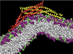
CRISPR-Cas9 conformational activation as elucidated from enhanced molecular simulationsProc. Natl. Acad. Sci. USA, Vol. 114 (28), 7260-7265 (2017)
CRISPR-Cas9 has become a facile genome editing technology, yet the structural and mechanistic features underlying its function are unclear. Here, we perform extensive molecular simulations in enhanced sampling regime, using a Gaussian accelerated Molecular Dynamics (GaMD) methodology, which probes displacements over hundreds of microseconds to milliseconds, to reveal the conformational dynamics of the endonuclease Cas9 during its activation toward catalysis. We disclose the conformational transition of Cas9 from its apo form to the RNA-bound form, suggesting a mechanism for RNA recruitment in which the domain relocations cause the formation of a positively charged cavity for nucleic acid binding. GaMD also reveals the conformation of a catalytically competent Cas9, which is prone for catalysis and whose experimental characterization is still limited. We show that, upon DNA binding, the conformational dynamics of the HNH domain triggers the formation of the active state, explaining how the HNH domain exerts a conformational control domain over DNA cleavage (Sternberg et al. Nature, 2015, 527, 110-113). These results provide atomic level information on the molecular mechanism of CRISPR-Cas9 that will inspire future experimental investigations aimed at fully clarifying the biophysics of this unique genome editing machinery and at developing new tools for nucleic acid manipulation based on CRISPR-Cas9.
CRISPR-Cas9 has become a facile genome editing technology, yet the structural and mechanistic features underlying its function are unclear. Here, we perform extensive molecular simulations in enhanced sampling regime, using a Gaussian accelerated Molecular Dynamics (GaMD) methodology, which probes displacements over hundreds of microseconds to milliseconds, to reveal the conformational dynamics of the endonuclease Cas9 during its activation toward catalysis. We disclose the conformational transition of Cas9 from its apo form to the RNA-bound form, suggesting a mechanism for RNA recruitment in which the domain relocations cause the formation of a positively charged cavity for nucleic acid binding. GaMD also reveals the conformation of a catalytically competent Cas9, which is prone for catalysis and whose experimental characterization is still limited. We show that, upon DNA binding, the conformational dynamics of the HNH domain triggers the formation of the active state, explaining how the HNH domain exerts a conformational control domain over DNA cleavage (Sternberg et al. Nature, 2015, 527, 110-113). These results provide atomic level information on the molecular mechanism of CRISPR-Cas9 that will inspire future experimental investigations aimed at fully clarifying the biophysics of this unique genome editing machinery and at developing new tools for nucleic acid manipulation based on CRISPR-Cas9.
Gaussian Accelerated Molecular Dynamics: Theory, Implementation and ApplicationsAnnual Reports in Computational Chemistry 13, 231-278 (2017).
A novel Gaussian Accelerated Molecular Dynamics (GaMD) method has been developed for simultaneous unconstrained enhanced sampling and free energy calculation of biomolecules. Without the need to set predefined reaction coordinates, GaMD enables unconstrained enhanced sampling of the biomolecules. Furthermore, by constructing a boost potential that follows a Gaussian distribution, accurate reweighting of GaMD simulations is achieved via cumulant expansion to the second order. The free energy profiles obtained from GaMD simulations allow us to identify distinct low energy states of the biomolecules and characterize biomolecular structural dynamics quantitatively. In this chapter, we present the theory of GaMD, its implementation in the widely used molecular dynamics software packages (AMBER and NAMD), and applications to the alanine dipeptide biomolecular model system, protein folding, biomolecular large-scale conformational transitions and biomolecular recognition.
A novel Gaussian Accelerated Molecular Dynamics (GaMD) method has been developed for simultaneous unconstrained enhanced sampling and free energy calculation of biomolecules. Without the need to set predefined reaction coordinates, GaMD enables unconstrained enhanced sampling of the biomolecules. Furthermore, by constructing a boost potential that follows a Gaussian distribution, accurate reweighting of GaMD simulations is achieved via cumulant expansion to the second order. The free energy profiles obtained from GaMD simulations allow us to identify distinct low energy states of the biomolecules and characterize biomolecular structural dynamics quantitatively. In this chapter, we present the theory of GaMD, its implementation in the widely used molecular dynamics software packages (AMBER and NAMD), and applications to the alanine dipeptide biomolecular model system, protein folding, biomolecular large-scale conformational transitions and biomolecular recognition.
‘MARTINIZING’ the Variational Implicit Solvent Model (VISM): Solvation Free Energy for Coarse-Grained ProteinsJ. Phys. Chem. 121 (27), pp 6538–6548 (2017)
Solvation is a fundamental driving force in many biological processes including biomolecular recognition and self-assembly, not to mention protein folding, dynamics, and function. The variational implicit solvent method (VISM) is a theoretical tool currently developed and optimized to estimate solvation free energies for systems of very complex topology, such as biomolecules. VISM’s theoretical framework makes it unique because it couples hydrophobic, van der Waals, and electrostatic interactions as a functional of the solvation interface. By minimizing this functional, VISM produces the solvation interface as an output of the theory. In this work, we push VISM to larger scale applications by combining it with coarse-grained solute Hamiltonians adapted from the MARTINI framework, a well-established mesoscale force field for modeling large-scale biomolecule assemblies. We show how MARTINI-VISM (MVISM) compares with atomistic VISM (AVISM) for a small set of proteins differing in size, shape, and charge distribution. We also demonstrate MVISM’s suitability to study the solvation properties of an interesting encounter complex, barnase–barstar. The promising results suggest that coarse-graining the protein with the MARTINI force field is indeed a valuable step to broaden VISM’s and MARTINI’s applications in the near future.
Solvation is a fundamental driving force in many biological processes including biomolecular recognition and self-assembly, not to mention protein folding, dynamics, and function. The variational implicit solvent method (VISM) is a theoretical tool currently developed and optimized to estimate solvation free energies for systems of very complex topology, such as biomolecules. VISM’s theoretical framework makes it unique because it couples hydrophobic, van der Waals, and electrostatic interactions as a functional of the solvation interface. By minimizing this functional, VISM produces the solvation interface as an output of the theory. In this work, we push VISM to larger scale applications by combining it with coarse-grained solute Hamiltonians adapted from the MARTINI framework, a well-established mesoscale force field for modeling large-scale biomolecule assemblies. We show how MARTINI-VISM (MVISM) compares with atomistic VISM (AVISM) for a small set of proteins differing in size, shape, and charge distribution. We also demonstrate MVISM’s suitability to study the solvation properties of an interesting encounter complex, barnase–barstar. The promising results suggest that coarse-graining the protein with the MARTINI force field is indeed a valuable step to broaden VISM’s and MARTINI’s applications in the near future.
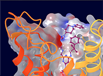
Protospacer Adjacent Motif-Induced Allostery Activates CRISPR-Cas9J. Amer. Chem. Soc., 139, 45, pp 16028-16031 (2017)
CRISPR-Cas9 is a genome editing technology with major impact in life sciences. In this system, the endonuclease Cas9 generates double strand breaks in DNA upon RNA-guided recognition of a complementary DNA sequence, which strictly requires the presence of a protospacer adjacent motif (PAM) next to the target site. Although PAM recognition is essential for cleavage, it is unknown whether and how PAM binding activates Cas9 for DNA cleavage at spatially distant sites. Here, we find evidence of a PAM-induced allosteric mechanism revealed by microsecond molecular dynamics simulations. PAM acts as an allosteric effector and triggers the interdependent conformational dynamics of the Cas9 catalytic domains (HNH and RuvC), responsible for concerted cleavage of the two DNA strands. Targeting such an allosteric mechanism should enable control of CRISPR-Cas9 functionality.
CRISPR-Cas9 is a genome editing technology with major impact in life sciences. In this system, the endonuclease Cas9 generates double strand breaks in DNA upon RNA-guided recognition of a complementary DNA sequence, which strictly requires the presence of a protospacer adjacent motif (PAM) next to the target site. Although PAM recognition is essential for cleavage, it is unknown whether and how PAM binding activates Cas9 for DNA cleavage at spatially distant sites. Here, we find evidence of a PAM-induced allosteric mechanism revealed by microsecond molecular dynamics simulations. PAM acts as an allosteric effector and triggers the interdependent conformational dynamics of the Cas9 catalytic domains (HNH and RuvC), responsible for concerted cleavage of the two DNA strands. Targeting such an allosteric mechanism should enable control of CRISPR-Cas9 functionality.
Manipulating Protein–Protein Interactions in Nonribosomal Peptide Synthetase Type II Peptidyl Carrier ProteinsBiochemistry, 56 (40), pp 5269–5273 (2017)
In an effort to elucidate and engineer interactions in type II nonribosomal peptide synthetases, we analyzed biomolecular recognition between the essential peptidyl carrier proteins and adenylation domains using nuclear magnetic resonance (NMR) spectroscopy, molecular dynamics, and mutational studies. Three peptidyl carrier proteins, PigG, PltL, and RedO, in addition to their cognate adenylation domains, PigI, PltF, and RedM, were investigated for their cross-species activity. Of the three peptidyl carrier proteins, only PigG showed substantial cross-pathway activity. Characterization of the novel NMR solution structure of holo-PigG and molecular dynamics simulations of holo-PltL and holo-PigG revealed differences in structures and dynamics of these carrier proteins. NMR titration experiments revealed perturbations of the chemical shifts of the loop 1 residues of these peptidyl carrier proteins upon their interaction with the adenylation domain. These experiments revealed a key region for the protein–protein interaction. Mutational studies supported the role of loop 1 in molecular recognition, as mutations to this region of the peptidyl carrier proteins significantly modulated their activities.
In an effort to elucidate and engineer interactions in type II nonribosomal peptide synthetases, we analyzed biomolecular recognition between the essential peptidyl carrier proteins and adenylation domains using nuclear magnetic resonance (NMR) spectroscopy, molecular dynamics, and mutational studies. Three peptidyl carrier proteins, PigG, PltL, and RedO, in addition to their cognate adenylation domains, PigI, PltF, and RedM, were investigated for their cross-species activity. Of the three peptidyl carrier proteins, only PigG showed substantial cross-pathway activity. Characterization of the novel NMR solution structure of holo-PigG and molecular dynamics simulations of holo-PltL and holo-PigG revealed differences in structures and dynamics of these carrier proteins. NMR titration experiments revealed perturbations of the chemical shifts of the loop 1 residues of these peptidyl carrier proteins upon their interaction with the adenylation domain. These experiments revealed a key region for the protein–protein interaction. Mutational studies supported the role of loop 1 in molecular recognition, as mutations to this region of the peptidyl carrier proteins significantly modulated their activities.
